The circulatory system and the heart's role in it ...
You may have often heard about the circulatory system in our body and
wondered what exactly it does. Today, we explain how it operates in
brief.
The circulatory system is responsible for the transport of water and
dissolved materials throughout the body, including oxygen, carbon
dioxide, nutrients, and waste. The circulatory system transports oxygen
from the lungs and nutrients from the digestive tract to every cell in
the body, allowing for the continuation of cell metabolism.
The circulatory system also transports the waste products of cell
metabolism to the lungs and kidneys where they can be expelled from the
body. Without this important function, toxic substances would quickly
build up in the body. The human circulatory system is organised into two
major circulations. Each has its own pump with both pumps being
incorporated into a single organ the heart.
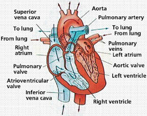
The two sides of the human heart are separated by partitions, the
interatrial septum and the interventricular septum. Both septa are
complete so that the two sides are anatomically and functionally
separate pumping units. The right side of the heart pumps blood through
the pulmonary circulation (the lungs) while the left side of the heart
pumps blood through the systemic circulation (the body).
You must be already knowing that the human heart is a specialised,
four-chambered muscle that maintains the blood flow in the circulatory
system. It lies immediately behind the sternum, or breastbone, and
between the lungs. The apex, or bottom of the heart, is tilted to the
left side.
The heart is made up of two muscle masses. One of these forms the two
atria (the upper chambers) of the heart, and the other forms the two
ventricles (the lower chambers). Both atria contract or relax at the
same time, as do both ventricles.
****
How the heart pumps blood
The heart muscle pumps the blood through the body by means of
rhythmical contractions (systole) and relaxations or dilations
(diastole). The heart's left and right halves work almost synchronously.
When the ventricles contract (systole), the valves between the atria
and the ventricles close as the result of increasing pressure, and the
valves to the pulmonary artery and the aorta open. When the ventricles
become flaccid during diastole, and the pressure decreases, the reverse
process takes place.
From the right atrium the blood passes to the right ventricle through
the tricuspid valve, which consists of three flaps (or cusps) of tissue.
The tricuspid valve remains open during diastole, or ventricular
filling. When the ventricle contracts, the valve closes, sealing the
opening and preventing backflow into the right atrium. Five cords
attached to small muscles, called papillary muscles, on the ventricles'
inner surface prevent the valves' flaps from being forced backward.
From the right ventricle, blood is pumped through the pulmonary or
semilunar valve, which has three half-moon-shaped flaps, into the
pulmonary artery.
This valve prevents backflow from the artery into the right
ventricle. From the pulmonary artery blood is pumped to the lungs where
it releases carbon dioxide and picks up oxygen.
****
The Systemic Circulation
From the lungs, the blood is returned to the heart through pulmonary
veins, two from each lung. From the pulmonary veins, the blood enters
the left atrium and then passes through the mitral valve to the left
ventricle.
As the ventricles contract, the mitral valve prevents backflow of
blood into the left atrium, and blood is driven through the aortic valve
into the aorta, the major artery that supplies blood to the entire body.
The aortic valve, like the pulmonary valve, has a semilunar shape.
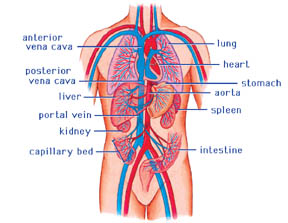
The aorta has many branches, which carry the blood to various parts
of the body. Each of these branches in turn has branches, and these
branches divide, and so on, until there are literally millions of small
blood vessels. The smallest of these on the arterial side of the
circulation are called arterioles.
They contain a great deal of smooth muscle, and because of their
ability to constrict or dilate, they play a major role in regulating
blood flow through the tissues.
The blood passing through the arterioles passes through a bed of
minute vessels called capillaries, which are a single cell thick. These
capillaries are so small that the red blood cells must line up single
file to pass through. The exchange of nutrients and waste products takes
place between the capillary blood and the tissue fluids. The
arterialised blood that enters the capillaries thus becomes venous blood
as it passes through them.
The capillaries empty the venous blood into collecting tubes called
venules, and these in turn empty into small veins, which empty into
larger veins, and so on, until finally all the blood returns to the
heart through two large veins, the superior and inferior vena cavae.
These terminate in the right atrium, and the systemic circulation is
complete.
A one-way flow of blood in this system is maintained by valves
located, not only in the heart, but in the veins as well. Some veins
also have semilunar valves and the pressure of contracting muscles
against the veins works with the action of these valves to increase the
venous return to the heart. This is the reason that exercise is so
important for the circulation.
*****
Coronary arteries and veins
The coronary arteries supply blood to the heart muscle. These vessels
originate from the aorta, immediately after the aortic valve, and branch
out through the heart muscle. The coronary veins transport the
deoxygenated blood from the heart muscle to the right atrium.
The heart's energy supply is almost completely dependent on these
coronary vessels. When the coronary vessels become blocked, as in
arteriosclerosis or hardening of the arteries, blood flow to the cardiac
muscle is compromised.
This is when the common "bypass surgery" is performed where the
coronary arteries are "bypassed" by replacing them with, for example, a
vein from the leg. A "double bypass" is when two coronary arteries are
bypassed. A "triple bypass" is when three are bypassed, etc. |