|
| Sunday, 15 January 2006 |
| Features |
| News Business Features Editorial
|
Health Guide
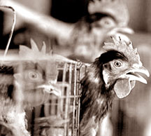
No bird flu in Sri Lanka by Dr. Keerthi Gunasekara
Aetiology Fowl plague was describe in 1878 as a serious disease of chickens in Italy. It was determined in 1955 that fowl plague (FP) virus is actually one of the influenza viruses. The AI viruses, along with the other influenza viruses, make up the virus family Orthomyxoviridae. The virus particle has an envelope with glycoprotein projections with haemagglutinating and neuraminidase activity. These two surface antigens, haemagglutinin (HA) and neuraminidase (NA), are the basis of describing the serologic identity of the influenza viruses using the letters H and N with the appropriate numbers in the virus designation e.g., H7N2. There are now 15 haemagglutinin and 9 neuraminidase antigens described among the Type A influenza viruses. The type designation (A, B, or C) is based upon the antigenic character of the M protein of the virus envelope and the nucleoprotein within the virus particle. All influenza viruses affecting domestic animals (equine, swine, avian) belong to Type A, and type A influenza virus is the most common type producing serious epidemics in humans. Types B and C do not affect domestic animals. Classical flow plague viruses have H7 as one of the surface antigens, but can have different N antigens. It was once believed that all H7 viruses are highly pathogenic fowl plague viruses, and that no other avian influenza viruses could produce a fowl-plague-like disease. When avirulent AI viruses with the H7 antigens were demonstrated in turkeys in 1971 and highly virulent AI viruses with the H5 antigen were first found in chickens in 1959, the necessity for redefining the term fowl plague or using other terminology became apparent. Because there are highly virulent AI viruses that possess H antigen other than the H7 and H5, AI viruses that do not produce clinical fowl plague, an international assembly of avian influenza specialists proposed that the term fowl plague no longer be used. They suggested that any AI virus, regardless of it is HA designation, meeting specified virulence requirements in the laboratory be designated highly pathogenic avian influenza (HPAI) and low pathogenic avian influenza (LPAI). Host range Most avian species appear to be susceptible to at least some of the AI viruses. A particular isolate may produce severe disease in turkeys but not in chickens or any other avian species. Therefore, it would be impossible to generalize on the host range for HPAI, for it will likely vary with the isolate. This assumption is supported by reports of farm outbreaks where only a single avian species of several species present on the farm became infected. Swine appear to be important in the epidemiology of infection of turkeys with swine influenza virus when they are in close proximity. Other mammals do not appear to be involved in the epidemiology of HPAI. The infection of humans with an H5 avian influenza virus in Hong Kong in 1997 has resulted in a reconsideration of the role of the avian species in the epidemiology of human influenza. Geographic distribution Highly pathogenic avian influenza viruses have periodically occurred in recent years in Australia (H7), England (H7), South Africa (H5), Scotland (H5), Ireland (H5), Mexico (H5), Pakistan (H7), and the United States (H5). Because laboratory facilities are not readily available in some parts of the world to differentiate Newcastle disease and HPAI, the actual incidence of HPAI in the world's poultry flocks is difficult to define. It can occur in any country, regardless of disease control measures, probably because of its prevalence in wild migratory waterfowl, sea birds and shore birds. Avian influenza has produced losses of variable severity, primarily in turkeys in the United States, since the mid-1960. The disease outbreaks in turkeys in the United States have been caused by AI viruses with many of the HA designations. It was in the fall of 1983 that a highly virulent H5 virus produced severe clinical disease and high mortality in chickens, turkeys, and guineafowl in Pennsylvania. This severe disease, is clinically indistinguishable from classical fowl. Incubation period The incubation period is usually 3 to 7 days, depending upon the isolate, the dose of inoculums, the species, and age of the bird. Clinical signs Infections of HPAI result in marked depression with ruffled feathers, inappetence, excessive thirst, cessation of egg production, and watery diarrhoea. Mature chickens frequently have swollen combs, wattles and edema surrounding the eyes. The combs are often cyanotic at the tips and may have plasma or blood vesicles on the surface with dark areas of ecchymotic haemorrhage and necrotic foci. The last eggs laid, after the onset of illness, are frequently without shells. The diarrhoea begins as watery bright green and progresses to almost totally white. Edema of the head, if present, is often accompanied by edema of the neck. The conjunctivae are congested and swollen with occasional haemorrhage. The legs, between the hocks and feet, may have areas of diffuse haemorrhage. Respiratory signs can be a significant feature of the disease, depending on the extent of tracheal involvement. Mucus accumulation can vary. It is not unusual in caged layers for the disease to begin in a localized area of the house and severely affect birds in only a few cages before it spreads to neighbouring cages. Death may occur within 24 hours of the first signs of disease, frequently within 48 hours, or be delayed for as long as a week. Some severely affected hens may occasionally recover.In broilers, the signs of disease are frequently less obvious with severe depression, inappetence, and a marked increase in mortality being the first abnormalities observed. Edema of the face and neck and neurological signs such as torticollis and ataxia may also be seen. To be continued Do you eat too fast? Most days, Mike Stepler wolfs down his lunch in less than 10 minutes. And most days, Stepler, a surveyor from Elsinboro, N.J., ends up with heartburn. Stepler wonders whether it was what he eats. Sometimes the lunch he chooses is very spicy. But his girlfriend thinks it has more to do with the pace at which the 20-year-old gobbles his food. His girlfriend may be on the right track. A recent study shows that, even among people who rarely experience heartburn, acid reflux - digestive juices moving backward from the stomach into the oesophagus - is more common if meals are hastily eaten. The study, published in the September issue of The American Journal of Gastroenterology, followed 20 healthy volunteers who were fed a meal, researchers thought was likely to trigger a little heartburn: a turkey burger, french fries and a Coke. "We had them come in on two different days," says study co-author Dr. Donald O. Castell, a professor of medicine and director of the oesophageal disorders program at Digestive Disease Center at the Medical University of South Carolina in Charleston. "And we randomly assigned them to eat the meal in either five minutes or in 30 minutes." So, people who ate in five minutes one day, ate in 30 the next and vice versa, Castell explains. Each of the volunteers was fitted with a probe that could detect acid reflux into the oesophagus. Researchers found that reflux was much more common when people who ate quickly. Castell says he and his colleagues decided to study fast eating after they noticed that medical students who gobbled down meals tended to have a lot of heartburn. "Over the years, many of them told us that they got indigestion if they ate too fast," Castell says. Food for thought So, what is it about fast eating that leads to reflux? If you eat too fast, your stomach fills up too quickly, says Dr. David Metz, a professor of medicine in the division of gastroenterology, director of the acid peptic disorders program and co-director of the GI physiology laboratory, all at the University of Pennsylvania in Philadelphia. When the upper part of the stomach stretches, it causes the vagus nerve to send a message to the brain, which then tells the lower oesophageal sphincter (LES) to relax, Metz explains. The LES is a mostly one-way valve that allows food to pass from the oesophagus into the stomach. When it opens the opposite way - from the stomach into the oesophagus - acid can seep up, leading to that familiar burning sensation in the chest that we know as heartburn. If you eat more slowly, the upper part of the stomach has more time to move the food along through the digestive tract, Metz says. The purpose of these transient relaxations of the LES is probably to allow people to belch when there's too much air in the stomach, Castell says. Creatures of habit What the study doesn't show, according to the experts, is whether slower eating would be a big benefit to people who regularly suffer heartburn. "We don't know whether it would be a 1 percent or a 40 percent contribution," says Dr. Bennett E. Roth, chief of clinical gastroenterology at the David Geffen School of Medicine at the UCLA. Besides, says Roth, the habit of bolting down food can be difficult to break. And these days, most people who come in with heartburn symptoms don't want to make drastic changes in the way they live, Roth says. "To be frank, they'd rather get a pill than have to turn their whole lives upside down with lifestyle modifications," he adds. Still, Metz says he'd like to see a study that looked at the impact of slower eating in people with frequent heartburn. But, even if it's proven that slow eating can relieve a lot of symptoms, that won't help people like Stepler, who says he only has about 10 minutes to eat lunch - which can range anywhere from three tacos to an 8-inch sub - when he's working out of the office. "I just eat and I'm on my way," Stepler says. |
|
| News | Business | Features
| Editorial | Security
| Produced by Lake House |


 Avian
influenza (AI) is a disease of viral aetiology that ranges from a mild or
even asymptomatic infection to an acute, fatal disease of chickens, turkeys,
guinea fowls, and other avian species, especially migratory waterfowl.
Avian
influenza (AI) is a disease of viral aetiology that ranges from a mild or
even asymptomatic infection to an acute, fatal disease of chickens, turkeys,
guinea fowls, and other avian species, especially migratory waterfowl.