|

Nuclear Medicine:
The key to accurate detection of illnesses
By Carol Aloysius
Despite its long history and claims by many scientists to have
discovered it, Nuclear Medicine remains shrouded in mystery even among
the medical community today.
As far back as 1896, Henri Becquerel, a French Physicist is said to
have discovered Nuclear Energy quite by accident when he left some X-ray
plates in storage next to uranium. But the discovery of Nuclear Medicine
itself is said to be attributed to the husband-wife team, Frederick
Jolinot ‘Curie and Irene Joiliot curie when in 1934, they discovered
artificially produced isotopes.
 |
|
Dr J.M Chandragupta
Udugama |
In Sri Lanka the Medical Faculty at Peradeniya had a Nuclear Medical
unit since 1978, followed by the Colombo Medical Faculty in 1990 and
subsequently the Lady Ridgeway Hospital and the Maharagama Cancer
Hospital.
The first private hospital to start a Nuclear Medicine Unit was the
Apollo Hospital in 2003.
So having been around for so long, why is this discipline, with its
well known far ranging benefits still a strange medical term to even
practicing doctors today?
The Sunday Observer spoke to the Consultant in Nuclear Medicine at
the Lanka Hospital (formerly known as Apollo Hospital) to find out why.
Dr J.M Chandragupta Udugama, a strong advocate of Nuclear Medicine
himself says he was unaware of the existence of this discipline in Sri
Lanka and learned about it quite by chance. He discusses what Nuclear
Medicine is and its potential for evaluating bone diseases, lung
functions, thyroid function and heart function as well as detection of
stage cancer and rare tumours of the pancreas and adrenal glands,
besides treating some of these debilitating diseases. As he points out,
“The procedure is painless and has no side effects. Even though it
involves using radioactive materials, the dose given is very low and
less harmful than an ordinary X-ray, or chemotherapy ”
Excerpts
Question: You have been a strong advocate of Nuclear Medicine
for a long time. How did you get interested in this specialised field?
As a student at Medical College or later as a practising doctor?
Answer: To be honest, I had no idea about Nuclear Medicine
while I was a medical student at the Colombo Medical Faculty. I got
familiar with the subject when I went to do my internship at the
Kurunegala Hospital. While there, I was trying to find out where I could
do a test to check the thyroid level of a patient and was informed that
this test was available at the Peradeniya Medical Faculty which had a
Nuclear Medicine Unit. I later became a medical officer at the
Peradeniya Nuclear Medicine Unit and developed a keen interest in the
subject, joining the Apollo Hospital in 2010 when I left government
service.
Q. Why are most doctors still unfamiliar with this discipline?
A. I believe it is because this subject is not given adequate
attention in our usual curriculum as medical students. Ideally we should
be introduced to Nuclear Medicine which is the medicine for the future,
in the Advanced Level classes.
Q. What is Nuclear Medicine?
 A. Nuclear Medicine is a medical speciality that uses small
amounts of radioactive materials (or tracers) to diagnose and treat a
variety of diseases. It is unique in that it diagnoses diseases based on
organ function and structure in contrast to diagnostic radiology which
determines presence of disease based on structural appearance. A. Nuclear Medicine is a medical speciality that uses small
amounts of radioactive materials (or tracers) to diagnose and treat a
variety of diseases. It is unique in that it diagnoses diseases based on
organ function and structure in contrast to diagnostic radiology which
determines presence of disease based on structural appearance.
Q. Under what discipline does it come?
A. Nuclear Medicine comes under the discipline of Imaging.
Basically, there are two branches of Imagining; 1) Radiology imaging and
2) Nuclear Imagining.
Q. What is the difference?
A. In Radiology imaging, the machines produce radiation e.g.
an X-ray where radiation goes through the patient’s body to give an
image of a particular site like the chest and bones. But in Nuclear
Medicine, you don’t use machines to produce radiation. The machine is
harmless by itself as it contains no radiation.
What happens is that you induce some form of radiation to the
patient, usually through the mouth, like drinking a solution of
radioactive iodine. This goes down the alimentary tract and digestive
tract and is absorbed by the thyroid cells.
Q. Isn’t that dangerous to the patient’s health?
A. In a Nuclear Medicine test (also known as a scan), very
small amounts of shortlived gamma emitting radioactive materials or
radiopharmaceuticals (tracers) are injected in, ingested by or inhaled
by the patient. As I said earlier it is less harmful than taking an
X-ray or going for a chemotherapy or radiation in the case of cancer
patients.
Q. You spoke about gamma emitting radioactive materials. What
exactly do you mean by this?
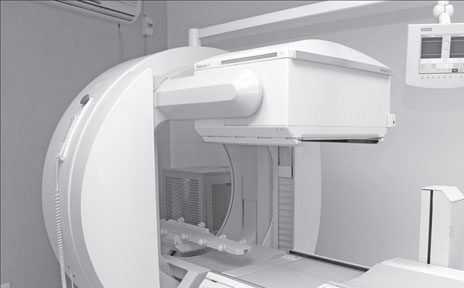
A. We use a special camera called the Gamma Camera to take
pictures of various organs of the body. The Gamma camera by detecting
traces in the organ, bone of tissues being imaged, records this
information on a computer screen or film, by using advanced software, we
are able to produce images of the site.
Q. What is the purpose of those images?
A. They are functional images which means they are only
visible if the organs where the camera points to are functioning.
How nuclear medicine helps children
It investigates abnormalities in the oesophagus, liver, gall bladder,
kidneys and intestines.
For women
Scintimammography
This is an important complement to mammography in patients with
suspected breast cancer. Nuclear medicine breast imaging is often used
for women who have particularly dense breast tissue, for women with
breast implants, or when multiple tumours are suspected. The technique
can also help differentiate between possible tumours and scar tissue
remaining from a previous mastectomy. |
Q. So a liver that is non functioning or a kidney that is non
functioning at the time the image is being taken will not be seen.
A. If a kidney has collapsed or not functioning, we will be
unable to get a clear picture or any picture.
Q. How does this image help the patient or the doctor treating
the patient?
A. By revealing the malfunctioning organ, the doctor or
surgeon will be able to administer the appropriate treatment.
Q. What kind of scans are commonly requested?
A. Most people ask for thyroid, kidney and bone scans since
their knowledge of the availability of other types of scans is less. So
these are more popular.
We also use different chemicals for different glands within the body
without altering the physiognomy of the system to produce the images.
However the most advanced techniques we use are in imaging the heart,
where we can see the blood distribution into the heart muscles, and
visualise the extent of the damage and the viability of the heart
muscles.
This test, by the way, is only available at this hospital.
Q. Does it require special training to perform the test?
A. Yes. Doctors and nurses have been specially trained for
this and in preparing patients undergoing the test.
Q. What do you see are the main benefits of Nuclear Medicine?
A. One of the biggest benefits is that the Nuclear Imaging
tests often identify abnormalities very early in the course of the
disease - long before some medical problems are apparent - clinically or
with other diagnostic tests.
For example, if a person gets a urine tract infection, it can develop
kidney granules or a scar. If you can detect the scar at the early
stage, you can prevent further damage to the kidney especially if it
were to start in childhood you can develop kidney complications if the
infection were not detected at its formative stages.
Q. Is the sky the limit for tests that can be done this way?
A. In a way yes. The Nuclear Medicine facility today, offers a
wide range of procedures. They include Whole body scans, Myocardial
Perfusion SPEC scans, dynamic renal scans for studying function and
drainage patterns of kidneys, DMSA, Renals scans for scars and
abnormalities, Hepatobiliary scans for gall bladder dysfunction, thyroid
scans, Parathyroid scans and Brain SPEC scans.
Q. Can these scans be done for people of any age?
A. Yes. They can be done on infants and elderly persons.
Q. Are there exceptions like pregnant women, for example,
since they are radioactive?
A. The only contraindication for Nuclear Medicine is
pregnancy. This is because the foetus is very vulnerable due to its
immature cells and radiation may harm it.
Q. Then what is the option open to a pregnant woman with a
kidney or liver problem?
A. Their condition will have to be managed with ultra sound
scans which don’t produce radiation till after delivery. Unless it is a
matter of life and death or absolutely necessary at the last stages of
pregnancy, will we subject pregnant women to a test or imaging using
radioactive rays.
Q. What about the effects on the unborn child if this happens?
A. We may have to do some Nuclear Medicine imaging very early
in life, but because we will be using very mild isotopes and tiny doses
it will be safe for the infant.
For example, Biliary Atrisea maldevelopment of the bile duct occurs
when bile doesn’t collect in the gall bladder resulting in liver damage.
So, gall bladder imaging is the only way we can test this condition
early, to help the surgeon to configurate the diagnosis and plan for
surgical correction.
Q. Can the ordinary man in the street afford these tests?
A. For various organs specific pharmacentives are imported of
the highest quality from England and Canada and the cardiac imaging
centre in the US because we don’t want to compromise the health of the
patient.
These medicines are produced by a handful of companies worldwide
which is another reason why they are expensive.
They are not in the open market but only available in limited stocks
in those manufacturing companies, because their shelf life is also very
short. That means you can’t stock large amounts. We have to import our
isotopes every fortnight.
Q. How are the tests performed?
A. Usually on an outpatient basis but also done on
hospitalised patients as well.
Q. How do you view the role of Nuclear Medicine in the future?
A. The new trend is not to just detect early but to treat
specific cancers such as colon cancer, ovarian cancer and pancreatic
cancer. To this end they have developed anti bodies and when combined
with radioisotopes and injected back into the systems, those anti bodies
get absorbed by the cancer cells and destroys them.
Adults suffer the effects of childhood bullying
A new study shows that serious illness, struggling to hold down a
regular job, and poor social relationships are just some of the adverse
outcomes in adulthood faced by those exposed to bullying in childhood.
It has long been acknowledged that bullying at a young age presents a
problem for schools, parents and public policy makers alike. Although
children spend more time with their peers than their parents, there is
relatively little published research on understanding the impact of
these interactions on their lives beyond school.
The results of the new study, published in Psychological Science,
highlight the extent to which the risk of problems related to health,
poverty, and social relationships are heightened by exposure to
bullying.
The study is notable because it looks into many factors that go
beyond health-related outcomes.
Psychological scientists Dieter Wolke and William E. Copeland led the
research team, looking beyond the study of victims and investigating the
impact on all those affected: the victims, the bullies themselves, and
those who fall into both categories, so-called “bully-victims.”
“We cannot continue to dismiss bullying as a harmless, almost
inevitable, part of growing up,” says Wolke. “We need to change this
mindset and acknowledge this as a serious problem for both the
individual and the country as a whole; the effects are long-lasting and
significant.”
The ‘bully-victims’ were at greatest risk for health problems in
adulthood, over six times more likely to be diagnosed with a serious
illness, smoke regularly, or develop a psychiatric disorder compared to
those not involved in bullying.
The results show that bully-victims are perhaps the most vulnerable
group of all. This group may turn to bullying after being bullied
themselves as they may lack the emotional regulation or support required
to cope with it.
“In the case of bully-victims, it shows how bullying can spread when
left untreated,” Wolke added.
“Some interventions are already available in schools but new tools
are needed to help health professionals to identify, monitor, and deal
with the ill-effects of bullying. The challenge we face now is
committing the time and resources to these interventions to try and put
an end to bullying.”
All the groups were more than twice as likely to have difficulty in
keeping a job, or committing to saving compared to those not involved in
bullying.
As such, they displayed a higher propensity for being impoverished in
young adulthood. However, the study revealed very few ill effects of
being the bully. After accounting for the influence of childhood
psychiatric problems and family hardships - which were prevalent among
bullies - the act of bullying itself didn't seem to have a negative
impact in adulthood.
“Bullies appear to be children with a prevailing antisocial tendency
who know how to get under the skin of others, with bully-victims taking
the role of their helpers,” said Wolke.
“It is important to finds ways of removing the need for these
children to bully others and, in doing so, protect the many children
suffering at the hand of bullies - they are the ones who are hindered
later in life.”
- MNT
Traffic pollution adversely affects asthma in adults
Asthma sufferers frequently exposed to heavy traffic pollution or
smoke from wood fire heaters, experienced a significant worsening of
symptoms, a new University of Melbourne led study has found.
The study is the first of its kind to assess the impact of traffic
pollution and wood smoke from heaters on middle-aged adults with asthma.
The results revealed adults who suffer asthma and were exposed to heavy
traffic pollution experienced an 80 percent increase in symptoms and
those exposed to wood smoke from wood fires experienced an 11 percent
increase in symptoms.
 Asthma affects more than 300 million people worldwide and is one of
the most chronic health conditions. Asthma affects more than 300 million people worldwide and is one of
the most chronic health conditions.
Dr John Burgess at the University of Melbourne said “it is now
recommended that adults who suffer asthma should not live on busy roads
and that the use of old wood heaters should be upgraded to newer
heaters, to ensure their health does not worsen.”
In the study, a cohort of 1,383 44-year old adults in the Tasmanian
Longitudinal Health Study were surveyed for their exposure to smoke from
wood fires and traffic pollution. Participants were asked to rate their
exposure.
The survey asked for exposure to the frequency of heavy traffic
vehicles near homes and the levels of ambient wood smoke in winter.
Results were based on the self-reporting of symptoms and the number
of flare-ups or exacerbations in a 12-month period. Participants
reported from between two to three flare-ups (called intermittent
asthma) to more than one flare-up per week (severe persistent asthma)
over the same time.
Traffic exhaust is thought to exacerbate asthma through airway
inflammation. Particles from heavy vehicles exhaust have been shown to
enhance allergic inflammatory responses in sensitised people who suffer
asthma.
“Our study also revealed a connection between the inhalation of wood
smoke exposure and asthma severity and that the use of wood for heating
is detrimental to health in communities such as Tasmania where use of
wood burning is common,” Dr Burgess said.
“Clean burning practices and the replacement of old polluting wood
stoves by new ones are likely to minimise both indoor and outdoor wood
smoke pollution and improve people's health,” he said.
“These findings may have particular importance in developing
countries where wood smoke exposure is likely to be high in rural
communities due to the use of wood for heating and cooking, and the
intensity of air pollution from vehicular traffic in larger cities is
significant.”
The study, published in Respirology , revealed no association between
traffic pollution and wood smoke and the onset of asthma.
- Medicalxpress
|


