|
| Sunday, 12 March 2006 |
| Junior Observer |
| News Business Features Editorial
|
Heart... The efficient pump Continued from last week Last week we discussed how the heart plays a key role in keeping you alive. Today we will check out some more interesting facts about the heart. Now you know that a human being's heart is about the size of that human being's fist. As the body develops, the heart grows at the same rate as the fist. So an infant's heart and fist are about the same size at birth.
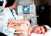
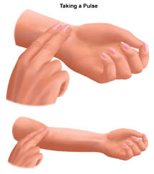
There are several phases of the foetal heart's development. At first, the heart is just a tube. It grows so fast that it needs more space, so it bends and twists back, forming the familiar shape. During the next phase, the two atria (chambers at the top) are partly separate, but there is just one big ventricle (chambers at the bottom). The next phase begins when the two atria are completely separate and the ventricles are just beginning to separate. Finally, the ventricles separate completely, and the heart is developed. During the foetal heart's stages of development, the heart actually takes on several distinct appearances. These heart structures resemble other animal hearts. During phase one, the tube-like heart is much like a fish heart. The second phase, with two chambers, resembles a frog heart. The three-chambered phase is similar to a snake or turtle heart. The final four-chambered heart structure distinguishes the human heart. The heart's structure makes it an efficient, never-ceasing pump. From the moment of development till the moment of death, the heart pumps. The heart, therefore, has to be strong. The average heart's muscle, called cardiac muscle, contracts and relaxes about 70 to 80 times per minute, without you ever having to think about it. As the cardiac muscle contracts, it pushes blood through the chambers and into the vessels. Nerves connected to the heart regulate the speed with which the muscle contracts. When you run, your heart pumps more quickly. When you sleep, your heart pumps more slowly. Considering how much work it has to do, the heart is surprisingly small. The average adult heart is about the size of a clenched fist and weighs about 11 ounces (310 grams) approximately. Located in the middle of the chest, behind the breastbone, between the lungs, the heart rests in a moistened chamber called the pericardial cavity, which is surrounded by the ribcage. The diaphragm, a tough layer of muscle, lies below. As a result, the heart is well protected. To monitor the heart, scientists can use x-ray or scanning technology to get a picture. To really explore the heart, scientists have to perform surgery. Heart surgery is very risky because the heart's pumping action is so critical for survival. If the heart stops pumping, the body cannot survive. Before beginning heart surgery, doctors connect the patient to a machine that pumps the blood for the heart. Only then is it safe for the doctor to stop the heart in order to operate. Because the heart is so well protected from outside danger, monitoring the heart can be a challenge. Medical scientists have several ways of "seeing" the heart without actually having to open the chest. Typically, open heart surgery is performed only as the last resort. To monitor the heart, a doctor begins by touching the chest. If the tip of the heart can be felt pushing against the chest, the heart may be enlarged. Thumping lightly on the chest gives an idea of the heart's shape. Your body's vital statistics also tell how well the heart is working. Next, the doctor places the stethoscope on the chest to hear the sound of the heart. The normal heart sounds, lub and dub, can be heard. Any unusual sounds can also be heard. Heart arrhythmias and murmurs are two possibilities. An arrhythmia is an irregular pattern of the heart's beat. A murmur indicates that blood is seeping through the closed valve separating the atrium and ventricle. An x-ray machine passes rays through the chest to make a shadow picture of the heart. The x-ray shows the size and position of the heart. It can also show any obvious deformities. Modern technology has provided even clearer pictures of the heart. Echocardiography and Electrocardiography are two techniques that provide detailed information about the heart without causing any real discomfort to the patient. Concluded Compiled by Chamitha Fact file * The heart is essentially a muscle. * The pumping of the heart is called the Cardiac Cycle, which occurs about 72 times per minute. This means that each cycle lasts about eight-tenths of a second. During this cycle, the entire heart actually rests for about four-tenths of a second. * The walls of the heart are made up of three layers, while the cavity is divided into four parts. There are two upper chambers, called the right and left atria, and two lower chambers, called the right and left ventricles. * A human being's heart is about the size of that human being's fist. As the body develops, the heart grows at the same rate as the fist. * The heart, like other body parts, needs oxygen in order to grow and develop properly. Throughout life, the heart needs only to be maintained and kept healthy, in order to function. If the heart ever ceases to pump blood, the body begins to shut down. Well, you know what happens when the body shuts down...you die. Check your pulse! When you go for a checkup, your doctor uses a stethoscope to listen carefully to your heart. A healthy heart makes a lub-dub sound with each beat. This sound comes from the valves shutting on the blood inside the heart.
The first sound (the lub) happens when the mitral and tricuspid valves close. The next sound (the dub) happens when the aortic and pulmonary valves close, after the blood has been squeezed out of the heart. You can find your pulse by lightly pressing on the skin anywhere there's a large artery running just beneath your skin. Two good places to find it are on the side of your neck and the inside of your wrist, just below the thumb. You'll know that you've found your pulse when you can feel a small beat
under your skin. Each beat is caused by the contraction (squeezing) of
your heart. If you want to find out what your heart rate is, use a watch
with a second hand, and count how many beats you feel in 1 minute. When
you are resting, you will probably feel between 70 and 100 beats per
minute. |
|
| News | Business | Features
| Editorial | Security
| Politics | Produced by Lake House |