|

'Near death' experience, a result of improved survival rates
Near death experience is the reported memory of all impressions
during a special state of consciousness, such as out of body experience,
pleasant feelings, seeing a tunnel, a light, deceased relatives or a
life review.
Psychologists defined the near death experience as the time of
clinical death where a patient is unconscious due to insufficient blood
supply to the brain because inadequate blood circulation, breathing or
both. In this situation, if resuscitation is not started within 5-10
minutes irreparable damage is done to the brain and the patient will
die.
Some people report near death experience (NDE) after a life
threatening crisis. A longitudinal study was conducted in Netherlands
between 2001-2009 which included 344 cardiac arrest patients who were
successfully resuscitated.
 The study was conducted among two groups, one group of cardiac
patients who had a cardiac arrest with a near death experience (62) or
18%, and in the other group of cardiac patients who did not have a near
death experience. This research studied the medical, pharmacological and
psychological experiences among the two groups. The occurrence of the
near death experience was not associated with duration of cardiac arrest
or unconsciousness, medication, or fear of death before cardiac arrest.
Significantly, in this study, more patients who had a near death
experience, especially deep experience, died with in 30 days after
resuscitation. The study was conducted among two groups, one group of cardiac
patients who had a cardiac arrest with a near death experience (62) or
18%, and in the other group of cardiac patients who did not have a near
death experience. This research studied the medical, pharmacological and
psychological experiences among the two groups. The occurrence of the
near death experience was not associated with duration of cardiac arrest
or unconsciousness, medication, or fear of death before cardiac arrest.
Significantly, in this study, more patients who had a near death
experience, especially deep experience, died with in 30 days after
resuscitation.
The researchers did not know, why so few cardiac patients reported
near death experience after resuscitation, but they thought it could be
explained by the age of the patients, memory power and brain damage
during the cardiac arrest some people who have survived a life
threatening crisis report an extraordinary experience.
Near death experience occurs with increasing frequency because of
improved survival rates resulting from modern techniques of
resuscitation.
The content of near death experience and the effects on patients seem
similar worldwide, across all cultures and times.
The subjective nature and absence of a frame of reference for this
experience lead to individual, cultural, and religious factors
determining the vocabulary used to describe and interpret the
experience.
Near death experiences are reported in many circumstances:
* Cardiac arrest in myocardial infarction (clinical death)
* Shock due to loss of blood in child birth or due to surgeries
Shock due to electrocution, allergies (anaphylactic shock) or shock
due to severe infections.
Attempted suicide, near drowning, serious traffic accidents,
mountaining accidents or in shipwreck situations.
Such experiences are also reported by people with severe depression
or without clear causes in fully concious people. Several theories on
the origin of near death experience have been proposed. Some
Psychologists think the experience is caused by changes in the brain,
such as brain cells dying due to lack of oxygen and nutrients. Another
theory is that the near death experience is due to changing state of
conciousness, in which perception (feelings), cognitive functioning,
emotion, and sense of identity, functioning independently from normal
body waking conciousness or dissociation from the body.
Patients who have undergone (survived) a near dear death experience
has transformational process in their lives so they do not have fear of
death and they accept any subsequent eventualities in a very rational
way.
Similar experiences to near-death can occur during the terminal phase
of an illness, and are called death-bed wishes.
Good short-term memory seems to be essential for remembering near
death experiences. Patients with memory defects after prolong
resuscitation reported fewer experiences.
Similar to near death experience can be induced through electrical
stimulation of some parts of the brain (temporal lobe and the hippo
campus of the brain), with high carbondioxide in the blood, during
training of fighter pilots (rapid acceleration when the brain does not
have enough oxygen). And in drug addicts (specially with LSD &
Ketamine). These induced experiences can consist of unconsciousness, out
of body experience, and seeing of light flashes of recollection from the
past.
These recollections, however, consist of fragmented and random
memories unlike the panoramic life-review that occurs in near death
experiences. Further, transformational processes with changing life
insight and disappearance of fear of death are not reported after
induced experiences.
The most important point to consider hear is that the clear
conciousness outside one's body the person experiencing at the moment,
when the brain no longer functions (flat EEG recording).
The same happens when there is cardiac arrest (flat ECG,) and when
there is flat EEG (brain is not functioning).
Furthermore, blind people (blind from birth), describes similar
out-of-body experience during cardiac arrest or when the brain is not
functioning (flat ECG & EEG).
More research should be conducted to explain scientifically the
phenomena of the near death experiences, It should be focused on
specific elements such as out-of-body experience and transcendence.
Dr. R.A. Ranjith Perera
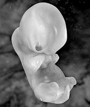
Scientists showcase how early human embryo acquires its shape
How is it that a disc-like cluster of cells transforms within the
first month of pregnancy into an elongated embryo? This mechanism is a
mystery that man has tried to unravel for millennia.
The first significant step towards understanding the issue was made
nearly a century ago in experiments conducted by the German
embryologists Hans Spemann and Hilde Mangold. The two used early newt
embryos and identified a group of cells within them which, upon
transplantation, formed a two-headed tadpole.
 In trying to understand why this happened, they concluded that what
occurred is that the transplanted cells organised the vicinity into
which they were placed to form a typical embryonic shape. They therefore
dubbed such cells "organizer" cells. In trying to understand why this happened, they concluded that what
occurred is that the transplanted cells organised the vicinity into
which they were placed to form a typical embryonic shape. They therefore
dubbed such cells "organizer" cells.
The new embryo possessed both its own organizers and the transplanted
ones, both of which organized nearby cells to form a head
structure.Recently, Israeli scientists from the Hebrew University of
Jerusalem have managed to generate human organiser cells, using human
embryonic stem cells. Based on the similarity that dominates the initial
developmental processes of all vertebrates, the group raised the human
cells in conditions which recapitulate those of early amphibian
embryogenesis. Within two days, the human cells started expressing genes
characteristic of the organizer cells.
To verify that these cells derived from human embryonic stem cells
posses a true organising ability, the researchers repeated Spemann and
Mangold's experiments.
Only this time, the human cells, rather than those of amphibians,
were transplanted into frog embryos.
The midline of an amphibian embryo is marked by a neural tube - a
tissue destined to form the embryo's central nervous system. To the
group's astonishment, some of the frog embryos that were transplanted
with the human cells possessed not one but two neural tubes. The second
tube was composed from frog cells, proving that the injected human cells
organized the cells in their vicinity to acquire a tubular shape.
Notes:
The research was conducted by Nadav Sharon, a graduate student under
the supervision of Nissim Benvenisty, the Hebert Cohn Professor of
Cancer Research at the Alexander Silberman Institute of Life Sciences at
the Hebrew University, in collaboration with Abraham Fainsod, the
Wolfson Family Professor of Genetics at the Hebrew University-Hadassah
Medical School, and was published in a recent issue of the Stem Cells
journal. Shape determination during human embryonic development is an
extremely important process, at which any aberration might lead to
miscarriage or the birth of a severely defected newborn.
The identification of the human organizer should allow better
understanding of this process.
Furthermore, the ability of the human organiser cells to shape a frog
neural tube may assist in forming human neural tubes in culture, from
which neural cells could be obtained for transplantation into people
with spinal damage, though much further research would be required to
reach that stage.
Source: Jerry Barach The Hebrew University of Jerusalem.
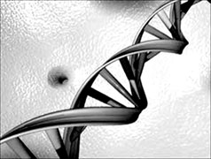
Molecular basis for DNA breakage, new approach to cancer treatment
Scientists have identified the molecular basis for DNA breakage, a
hallmark of cancer cells. The findings of this research have been
published in the journal Molecular Cell.
The DNA encodes the entire genetic information required for building
the proteins of the cell. Hence, DNA breaks disrupt the proteins and
lead to changes in the cell function. These changes can lead to defects
in the control of cellular proliferation resulting in cancer
development.
 Using cutting edge technologies, researchers Prof. Batsheva Kerem and
doctoral student Efrat Ozeri-Galai, of the Alexander Silverman Institute
of Life Sciences in the Faculty of Science were able to characterize for
the first time the DNA regions which are the most sensitive regions to
breakage in early stages of cancer development. Using cutting edge technologies, researchers Prof. Batsheva Kerem and
doctoral student Efrat Ozeri-Galai, of the Alexander Silverman Institute
of Life Sciences in the Faculty of Science were able to characterize for
the first time the DNA regions which are the most sensitive regions to
breakage in early stages of cancer development.
This is a breakthrough in our understanding of the effect of the DNA
sequence and structure on its replication and stability.
"A hallmark of most human cancers is accumulation of damage in the
DNA, which drives cancer development," says Prof. Kerem. "In the early
stages of cancer development, the cells are forced to proliferate. In
each cycle of proliferation the DNA is replicated to ensure that the
daughter cells have a full DNA. However, in these early stages the
conditions for DNA replication are perturbed, leading to DNA breaks,
which occur specifically in regions defined as 'fragile sites'."
In this research Prof. Kerem and Ozeri-Galai used a sophisticated new
methodology which enables the study of single DNA molecules, in order to
study the basis for the specific sensitivity of the fragile sites.
The findings are highly important since they shed new light on the
DNA features and on the regulation of DNA replication along the first
regions that break in cancer development.
The results show that along the fragile region there are sites that
slow the DNA replication and even stop it. In order to allow completion
of the DNA replication the cells activate already under normal
conditions mechanisms that are usually used under stress. As a result,
under conditions of replication stress, such as in early cancer
development stages, the cell has no more tools to overcome the stress,
and the DNA breaks.
The results of this study reveal the molecular mechanism that
promotes cancer development. Currently, different studies focus on the
very early stages of cancer development aiming to identify the events
leading to cancer on the one hand and on their inhibition, on the other.
The result of the current research identified for the first time DNA
features that regulate DNA replication along the fragile sites, in early
stages of cancer development.
In the future, these findings could lead to the development of new
therapeutic approaches to restrain and/or treat cancer.
Source: Jerry Barach The Hebrew University of Jerusalem
Greater risk of relapse in patients using anti-depressants
Patients who use anti-depressants are much more likely to suffer
relapses of major depression than those who use no medication at all,
concludes a McMaster researcher.
In a paper that is likely to ignite new controversy in the hotly
debated field of depression and medication, evolutionary psychologist
Paul Andrews concludes that patients who have used anti-depressant
medications can be nearly twice as susceptible to future episodes of
major depression.
 The meta-analysis suggests that people who have not been taking any
medication are at a 25 per cent risk of relapse, compared to 42 per cent
or higher for those who have taken and gone off an anti-depressant. The meta-analysis suggests that people who have not been taking any
medication are at a 25 per cent risk of relapse, compared to 42 per cent
or higher for those who have taken and gone off an anti-depressant.
Andrews and his colleagues studied dozens of previously published
studies to compare outcomes for patients who used anti-depressants
compared to those who used placebos.
They analysed research on subjects who started on medications and
were switched to placebos, subjects who were administered placebos
throughout their treatment, and subjects who continued to take
medication throughout their course of treatment. Andrews says
anti-depressants interfere with the brain's natural self-regulation of
serotonin and other neurotransmitters, and that the brain can
overcorrect once medication is suspended, triggering new depression.
Though there are several forms of anti-depressants, all of them
disturb the brain's natural regulatory mechanisms, which he compares to
putting a weight on a spring.
The brain, like the spring, pushes back against the weight. Going off
antidepressant drugs is like removing the weight from the spring,
leaving the person at increased risk of depression when the brain, like
the compressed spring, shoots out before retracting to its resting
state.
"We found that the more these drugs affect serotonin and other
neurotransmitters in your brain - and that's what they're supposed to do
- the greater your risk of relapse once you stop taking them," Andrews
says. "All these drugs do reduce symptoms, probably to some degree, in
the short-term. The trick is what happens in the long term.
Our results suggest that when you try to go off the drugs, depression
will bounce back. This can leave people stuck in a cycle where they need
to keep taking anti-depressants to prevent a return of symptoms."
Andrews believes depression may actually be a natural and beneficial -
though painful - state in which the brain is working to cope with
stress.
"There's a lot of debate about whether or not depression is truly a
disorder, as most clinicians and the majority of the psychiatric
establishment believe, or whether it's an evolved adaptation that does
something useful," he says. Longitudinal studies cited in the paper show
that more than 40 per cent of the population may experience major
depression at some point in their lives.
Most depressive episodes are triggered by traumatic events such as
the death of a loved one, the end of a relationship or the loss of a
job.
Andrews says the brain may blunt other functions such as appetite,
sex drive, sleep and social connectivity, to focus its effort on coping
with the traumatic event. Just as the body uses fever to fight
infection, he believes the brain may also be using depression to fight
unusual stress. Not every case is the same, and severe cases can reach
the point where they are clearly not beneficial, he emphasises.
Source: Wade Hemsworth McMaster University
Genes vital in preventing childhood leukaemia identified
Researchers have identified genes that may be important for
preventing childhood leukaemia. Acute lymphoblastic leukaemia (ALL) is a
cancer of the blood that occurs primarily in young children. It's
frequently associated with mutations or chromosomal abnormalities that
arise during embryonic or fetal development. Working with mice,
researchers led by Rodney DeKoter identified two key genes that appear
essential in the prevention of B cell ALL, the most common form of ALL
in children. The study is published online in Blood, the Journal of the
American Society of Haematology.
In the study, mice were generated with mutations in two genes called
PU.1 and Spi-B. Mutation of either PU.1 or Spi-B individually had little
effect. Unexpectedly, mutation of both genes resulted in 100% of the
mice developing B cell ALL. Eighty percent of ALL cases in children are
of the B cell type. The study found PU.1 and Spi-B have unanticipated
functional redundancy as "tumor suppressor" genes that prevent
leukaemia. "You can think of PU.1 and Spi-B proteins as brakes on a car.
If the main brake (PU.1) fails, you still have the emergency brake
(Spi-B). However, if both sets of brakes fail, the car speeds out of
control," explains DeKoter, an associate professor in the Department of
Microbiology & Immunology at Western's Schulich School of Medicine &
Dentistry. "And uncontrolled cell division is an important cause of
leukaemia."
PU.1 is an essential regulator in the development of the immune
system, and mutations in this gene have been previously associated with
human ALL. DeKoter hopes these studies will ultimately lead to improved,
less toxic, therapies for childhood leukaemia. Currently, about 80% of
ALL patients go into complete remission when treated with aggressive
chemotherapy. DeKoter is also affiliated with the Centre for Human
Immunology at Western and the Children's Health Research Institute. The
lead author on the paper is Kristen Sokalski, a 2011 BMSc graduate with
an honours specialization in Biochemistry of Infection & Immunity.
Stephen Li and Marek Gruca, both MSc students supervised by DeKoter, and
Ian Welch and Heather Cadieux-Pitre of Western's Veterinary Services
also worked on the project.
Source: Kathy Wallis University of Western Ontario
|


")


