|

Threat of Dengue Fever
Greater in rural areas than in cities:
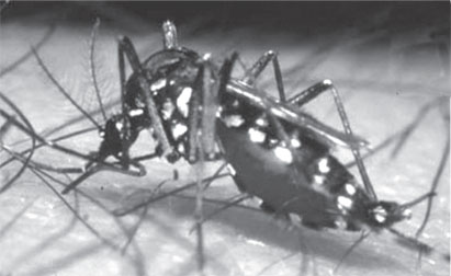 In dengue-endemic areas such as South-East Asia, in contrast to
conventional thinking, rural areas rather than cities may bear the
highest burden of dengue fever - a viral infection that causes sudden
high fever, severe headache, and muscle and joint pains, and can lead to
a life-threatening condition, dengue hemorrhagic fever. In dengue-endemic areas such as South-East Asia, in contrast to
conventional thinking, rural areas rather than cities may bear the
highest burden of dengue fever - a viral infection that causes sudden
high fever, severe headache, and muscle and joint pains, and can lead to
a life-threatening condition, dengue hemorrhagic fever.
In a study from the Nagasaki Institute of Tropical Medicine, Japan,
the authors analysed a population in Kanh-Hoa Province in south-central
Vietnam (~350,000 people) that was affected by two dengue epidemics
between January 2005 and June 2008.
They found that at low human population densities, mostly in rural
areas, dengue risk is up to three times higher than in cities,
presumably because the number of mosquitoes per individual is higher in
low density areas.
The authors show that severe outbreaks of dengue occur almost
exclusively in areas falling within a narrow range of human population
densities with limited access to tap water, where water storage vessels
provide breeding sites for the mosquitoes causing dengue fever. However,
as the actual number of people who contract dengue fever in populated
areas is high, urban areas still substantially contribute to dengue
epidemics.
The authors argue that improving water supply and vector control in
areas with a human population density critical for dengue transmission
could increase the efficiency of control efforts.
The authors say: “Ideally, all people should have access to reliable
tap water, not only to reduce the burden of dengue but also a range of
other diseases associated with inadequate water supply such as diarrhoea
or trachoma, and to realize important economic benefits.” However as
supplying everyone with tap water is not a realistic short-term option
in many low-income settings, reducing mosquito breeding around human
settlements is an uphill struggle.
The authors conclude: “Additional intervention measures in areas with
a human population density critical for dengue virus transmission could
increase the efficiency of vector control, especially since population
density figures are relatively easy to obtain.”
Courtesy: Medical News
Opticians could enable early identification of diabetes with a
simple blood test
A simple finger prick test during routine eye examinations at high
street opticians could help to identify millions of people with
previously undiagnosed Type 2 diabetes, according to new research.
 The researchers suggest earlier diagnosis could set people on the
road to better management of the disease, which is the leading cause of
blindness in the working age population, and that this could ultimately
result in cost-savings for the NHS. The researchers suggest earlier diagnosis could set people on the
road to better management of the disease, which is the leading cause of
blindness in the working age population, and that this could ultimately
result in cost-savings for the NHS.
The Durham University study suggests that screening for the condition
in unconventional settings, such as opticians, chiropodists or dentists,
could find those people who would not routinely visit their GP, and
could have potential worldwide.
It is estimated that 150 million people worldwide have diabetes but
up to 50 per cent of people who have the condition are thought to be
undetected and may only be diagnosed when complications occur.
It has already been shown that pharmacies and chiropodists have the
capacity to carry out simple blood tests to identify Type 2 diabetes.
The researchers say other places such as dentists could potentially be
used to offer the test.
The pilot study, carried out by Durham University and The James Cook
University Hospital in Middlesbrough, focused on opticians and the
findings are published in the British Journal of General Practice.
It found that out of 1,000 people visiting their opticians for an eye
test who were found to have one or more risk factors of diabetes, such
as increased body mass index or aged over 40, almost 32 per cent were
referred to their GP for further investigation after having their blood
glucose levels checked.
The researchers say high street opticians are an under-utilised
resource in the efforts to identify the large numbers of people with
undiagnosed diabetes.
Currently, most screening for diabetes is carried out in medical
settings, mostly by family doctors, but there are many people who do not
visit their GP for preventative care, even if they are in an at-risk
group.
While optometrists have an established role in screening people with
known diabetes for eye disease, they are presently not involved in
identifying diabetes.
Dr. Jenny Howse from Durham University’s School of Medicine and
Health, said: “Charities’ campaigns have managed to reduce the
proportion of people with undiagnosed diabetes but there is still a
‘hard-to-reach’ group who remain undiagnosed.
Opticians could provide routine, non-emergency care and the simple
screening can be done outside usual medical settings, such as GP
surgeries.” In the study, which involved five high street optometry
practices, people were asked a number of questions to identify any
diabetes risk factors.
If they had one or more, optical assistants conducted a simple finger
prick test, called a random capillary blood glucose (rCBG) test, to
assess the blood glucose levels. In keeping with current Royal
Pharmaceutical Society and Diabetes UK guidelines for screening in
pharmacies, those with raised blood glucose levels were advised to visit
their GP for further investigations.
Dr Howse said: “The screening test is less invasive and time
consuming than fasting blood glucose and oral glucose tolerance tests.
“Already pharmacists and chiropodists have shown it is feasible to offer
screening in their practices, here in the UK as well as in Australia and
Switzerland. In the US, 60 per cent of adults visit dentists at least
once a year for standard check-ups and those practices could be suitable
locations to screen for diabetes.
“In the UK, our initial results show screening for diabetes in
opticians is a feasible option but we now need to look at the
practicalities of delivering it, including liaison between opticians and
GPs and the time costs for opticians.” Type 2 diabetes is the most
common type of diabetes and the risk of developing it increases as you
get older.
It develops when the body does not produce enough insulin to maintain
a normal blood glucose level, or when the body is unable to effectively
use the insulin that is being produced.
Genetic factor implicated in heartbeat defect
Gene regulation can make hearts beat out of sync, offering new hope
for the millions who suffer from a potentially fatal heart condition. In
a paper being published Gladstone Investigator Benoit G. Bruneau,
announces the identity of the molecular regulator that uses electrical
impulses to synchronize each heartbeat.
 Abnormalities in heartbeat synchronization, called heart arrhythmias,
are a cause of death for the 5.7 million Americans who suffer from heart
failure, a condition in which the heart can’t pump enough blood to meet
the body’s needs. Abnormalities in heartbeat synchronization, called heart arrhythmias,
are a cause of death for the 5.7 million Americans who suffer from heart
failure, a condition in which the heart can’t pump enough blood to meet
the body’s needs.
At least 300,000 people die of heart failure each year in the United
States alone.“This is important progress for a better understanding of
heart arrhythmias, which when combined with heart failure can be fatal,”
said Deepak Srivastava, MD, who directs all cardiovascular research at
Gladstone. “This is the first published research about a genetic
regulator that coordinates the timing of the electrical impulses that
make the heart beat properly.”
In many animals, including humans, electrical impulses must spread
rapidly and in a coordinated fashion along a dedicated network of
cardiac cells in order for the heart to pump blood efficiently to the
rest of the body. A genetic regulator, called Irx3, coordinates these
impulses.
When Dr. Bruneau and his team switched off the Irx3 gene in mice, the
heart’s pumping fell out of sync. The electrical impulses- which
normally follow a rapid path throughout the heart - diffused slowly and
had trouble reaching their intended destinations. The mice developed
arrhythmias as the heart’s chambers lost the capacity to beat in time.
Dr. Bruneau, conducted the research in collaboration with two
Canadian labs. “These findings have potential implications for the
prevention and treatment of human heart disease, once we better
understand Irx3’s role in the human heart,” said Dr. Bruneau.
“An important avenue to explore could be whether humans with
arrhythmias have mutations in the Irx3 gene.”
Courtesy: Gladstone Institutes
What’s behind hypertension?
 Each day we consume liquids in order to keep hydrated and maintain
our body’s fluid balance. But just as a water balloon can get overtaxed
by too much liquid, the human body is negatively affected when it
retains fluids because it is unable to eliminate them properly. Each day we consume liquids in order to keep hydrated and maintain
our body’s fluid balance. But just as a water balloon can get overtaxed
by too much liquid, the human body is negatively affected when it
retains fluids because it is unable to eliminate them properly.
One of the key variables influencing how much fluid we hold in our
bodies is ordinary table salt (sodium chloride). The consequences of
excess fluid retention can be severe, causing not only edema (excess of
body fluid), but also high blood pressure (hypertension), which the
Centers for Disease Control estimates affects nearly one in three
American adults and last year carried an estimated financial toll of
$76.6 billion for the period.
What is the connection between fluid balance and hypertension? The
7th International Symposium on Aldosterone and the ENaC/Degenerin Family
of Ion Channels, sponsored by the American Physiological Society,
explores this public health concern in detail. New scientific findings,
coupled with talks by experts from around the world working in
aldosterone and epithelial sodium channel (ENaC) research, is offering
insight on the effect these substances have on blood pressure, the
cardiovascular system and other organ systems.
Aldosterone and ENaC can affect fluid regulation in several ways.
According to Dr. David Pearce, “Aldosterone controls ENaC, the key
sodium-transporting protein in the kidney tubule cells. It stimulates
the amount of sodium reabsorbed by the body as regulated by ENaC,” he
said. “The more sodium and water there is in the body, the more
circulating fluid there is for the heart to contend with. When the
process goes wrong, it can result in high blood pressure.”
Source: American Physiological Society -APS
Study finds increased light may moderate fearful reactions
Biologists and psychologists know that light affects mood, but a new
study indicates that light may also play a role in modulating fear and
anxiety.
Psychologist Brian Wiltgen and Biologists Ignacio Provencio and
Daniel Warthen of U.Va.’s College of Arts & Sciences worked together to
combine studies of fear with research on how light affects physiology
and behaviour.
Using mice as models, they learned that intense light enhances fear
or anxiety in mice, which are nocturnal, in much the same way that
darkness can intensify fear or anxiety in diurnal humans.
The finding is published in the Aug. 1 issue of the journal
Proceedings of the National Academy of Sciences.
“We looked at the effect of light on learned fear, because light is a
pervasive feature of the environment that has profound effects on
behaviour and physiology,” said Wiltgen, an assistant professor of
psychology and an expert on learning. “Light plays an important role in
modulating heart rate, circadian rhythms, sleep/wake cycles, digestion,
hormones, mood and other processes of the body. In our study we wanted
to see how it affects learned fear.”
Fear is a natural mechanism for survival. Some fears such as of loud
noise, sudden movements and heights appear to be innate. Humans and
other mammals also learn from their experiences, which include dangerous
or bad situations. This “learned fear” can protect us from dangers.
That fear also can become abnormally enhanced in some cases,
sometimes leading to debilitating phobias. About 40 million people in
the United States suffer from dysregulated fear and heightened states of
anxiety. “Studies show that light influences learning, memory and
anxiety,” Wiltgen said. “We have now shown that light also can modulate
conditioned fear responses.”
“In this work we describe the modulation of learned fear by ambient
light,” said Provencio, an expert on light and photoreception. “The
dysregulation of fear is an important component of many disorders,
including generalized anxiety disorder, panic disorder, specific phobias
and post-traumatic stress disorder. Understanding how light regulates
learned fear may inform therapies aimed at treating some of these
fear-based disorders.”
The researchers used a common method for studying learned fear. They
cued their mice with a minute-long tone that was followed two seconds
later by a quick, mild electrical shock.
The mice learned to associate the tone with the shock and quickly
became conditioned to duck down and remain motionless when they heard
the tone, in the same way they would if a predator appeared.
The researchers discovered that by intensifying the ambient light,
the mice showed a greater fear reaction to the tone than when the light
was dimmer. This makes sense Wiltgen said, because mice naturally avoid
detection by predators by hunkering down motionless as a defence
mechanism.
In a natural habitat they likewise would become particularly anxious
in the presence of a predator in bright light where they would be more
easily detected.
“We showed that light itself does not necessarily enhance fear, but
more light enhances learned fear,” Wiltgen said. “It may be similar to a
person learning to be more fearful in the dark.”The researchers wanted
to understand what visual pathways to the brain in mammals may be
responsible for this behaviour in the presence of more light.
The eye has two pathways that begin in the retina and end in the
brain: one is image-forming and made up of rods and cones; the other is
the non-image-forming retinal ganglion cells where melanopsin, a
circadian rhythm-regulating photo-pigment, is located.
Source: University of Virginia
How impulsiveness is controlled by the brain
How the brain controls impulsive behaviour may be significantly
different than psychologists have thought for the past 40 years.
That is the unexpected conclusion of a study by an international team
of neuroscientists.
Impulse control is an important aspect of the brain’s executive
functions - the procedures that it uses to control its own activity.
Problems with impulse control are involved in ADHD and a number of other
psychiatric disorders including schizophrenia. The current research set
out to better understand how the brain is wired to control impulsive
behaviour.
“Our study was focused on the control of eye movements, but we think
it is widely applicable,” said Vanderbilt Ingram Professor of
Neuroscience Jeffrey Schall, co-author of the new study.
Understanding impulse control
There are two sets of neurons that control how we process and react
to what we see, hear, smell, taste or touch. The first set, sensory
neurons, respond to different types of stimuli in the environment. They
are connected to movement neurons that trigger an action when the
information they receive from the sensory neurons reaches a certain
threshold. Response time to stimuli varies considerably depending on a
number of factors. When accuracy is important, for example, response
times lengthen. When speed is important, response times shorten.
According to Logan, there is clear evidence of a link between
reaction time variations and certain mental disorders. “In
countermanding tests, the response times of people with ADHD don’t slow
down as much following a stop-signal trial as normal subjects, while
response times of schizophrenics tend to be much slower than normal,” he
said.
Since the 1970’s, researchers have believed that the brain controls
these response times by altering the threshold at which the movement
neurons trigger an action: When rapid action is preferable, the
threshold is lowered and when greater deliberation is called for, the
threshold is increased.
In a direct test of this theory, however, Logan, Palmeri, Schall and
their collaborators found that differences in when the movement neurons
began accumulating information from the sensory neurons - rather than
differences in the threshold - appear to explain the adjustment in
response times.
This discovery forced them to make major modifications in the
existing cognitive model of impulse control and is an example of the
growing usefulness of such models to understand in much greater detail
what is occurring in the brain to cause both normal and abnormal
behaviours.
“Psychopathologists are beginning to use these models to make
connections with various brain disorders that we haven’t been able to
make before,” Palmeri said.
In the experiment
In the experiment, the delay between the appearance of the target and
stop signals ranged from 25 milliseconds to 275 milliseconds. During
this time, the movement neurons are still processing the signals
generated by the appearance of the target.
The longer the delay, the more difficult it is for the monkey to keep
from glancing at the target. In both humans and monkeys, the reaction
time in these tasks is significantly longer immediately following the
stop signal.
The researchers believe their discovery is significant because it
sheds new light on how the brain controls all sorts of basic impulses.
It is possible that neurons from the medial frontal cortex, which
performs executive control of decision-making, in the parietal lobe,
which determines our spatial sense, or the temporal lobe, which plays a
role in memory formation, may affect impulse control by altering the
onset delay time of neurons involved in a number of other basic
stimulus/response reactions. The project was supported by awards and
grants from the National Institutes of Health, the National Science
Foundation, the Canadian Institute of Health Research, the Ontario
Ministry of Research and Innovation and the ELJB Foundation.
- MNT
|


")

